Quantum technology for cancer imaging
Applied quantum mechanics in medicine
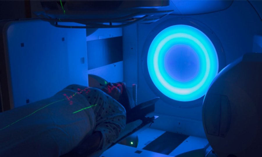
MUNICH: Tracing the metabolism of tumor cells using magnetic resonance imaging (MRI) has not been feasible in routine clinical settings hitherto. Now, an interdisciplinary research team including the Technical University of Munich (TUM) is working to advance the development of a quantum-based hyperpolarizer so that it can be deployed in clinical applications. The goal is to significantly improve MRI imaging of metabolic processes – for example, to allow earlier and more accurate assessment of tumors, as well as to improve the selection and monitoring of tumor therapies.
Quantum mechanics describes physical phenomena at the smallest of scales – in the domain of molecules, atoms, atomic nuclei, and even smaller units. The drive to revolutionize diverse domains of our daily lives using quantum technology like quantum computing or quantum sensors surfaced well before this year's Nobel Prize in Physics was awarded to three scientists for their work in this field. How can these new technologies be deployed in the field of medicine?
Metabolic imaging makes metabolic processes visible
Detecting cancer cells in early stages, assessing them with greater precision and evaluating the effectiveness of treatments faster is facilitated by the visualization of metabolic processes in both diseased and healthy cells. This is known as metabolic imaging. To this end, diagnostically relevant molecules are injected into the body and their metabolism is monitored.
One approach is to use positron emission tomography (PET). However, this method requires radioactive substances and cannot distinguish between the initial and end products in metabolic processes. Magnetic resonance imaging (MRI), on the other hand, allows metabolic imaging of various metabolites without using radioactive substances. Albeit only if the MRI signal of the injected molecules is amplified sufficiently to make it detectable. Although initial patient studies show great potential of metabolic imaging with MRI, the signal amplification technologies deployed up to now are prohibitively expensive, insufficiently robust, or slow. This has prevented routine deployment of these technologies in clinical settings up to now.
The interdisciplinary research team of the "Revolutionizing Cancer Imaging with Quantum Technologies" project (QuE-MRI) is now developing a new solution: A so-called quantum hyperpolarizer uses quantum physical laws to amplify the signal of metabolic molecules in the MRI up to 100,000-fold.
Imaging with the laws of quantum mechanics
The technology of common MRI machines takes advantage of quantum mechanical properties of atomic nuclei associated with the so-called spin, or angular momentum. Each nuclear spin generates a magnetic moment, not unlike the dipole magnet of a compass needle.
The alignment of the nuclear spins determines the strength of the overall magnetic moment of the atomic nuclei. This in turn determines the signal strength, which is used for magnetic resonance imaging. When the directional distribution of the magnetic moments is random, they cancel each other out and the MRI machine detects no signal. The strongest signal is achieved when the magnetic moments of the nuclear spins point in the same direction, resulting in the maximum effective magnetization.
MRI uses very strong magnetic fields to make this possible. Nonetheless, the magnetic moments of the nuclear spins are nearly randomly distributed and thus have only low effective magnetization. The technique of hyperpolarization boosts the effective magnetization of the nuclear spins by factor of 10,000 to 100,000, thereby significantly increasing the sensitivity of MRI.
Hyperpolarization of diagnostically relevant metabolic molecules
However, in practice enticing the atomic nuclei of the metabolic molecules into a hyperpolarized state is difficult. The researchers therefore use an intermediate step based on a special magnetic state of hydrogen, called para-hydrogen. This can be produced at low temperatures using known methods with liquid nitrogen and stored in gas cylinders.
The properties of para-hydrogen also build on the laws of quantum mechanics. While para-hydrogen itself is magnetically shielded and not measurable using magnetic resonance methods, its spin configuration can hyperpolarize other atomic nuclei, increasing their visibility in MRI.
Using this approach, the researchers hyperpolarize molecules important for studying metabolic processes. Pyruvate, for example, a metabolic product that is processed into lactic acid by tumors, is particularly suitable for diagnostic purposes. The researchers dock para-hydrogen onto pyruvate in the hyperpolarizer and use its spin configuration to hyperpolarize a carbon atom of pyruvate in a magnetic field using radio waves. The signal from pyruvate is thereby enhanced in MRI, allowing the corresponding metabolic process to be visualized with temporal resolution.
Project partners have already developed functional prototypes of the hyperpolarizer. In the QuE-MRI project, researchers, physicians, industrial partners, and developers in the fields of medicine, physics, chemistry and engineering are now collaborating closely to optimize these prototypes so that the hyperpolarizer can be deployed clinically on a large scale. In addition, the project team plans to validate the non-invasive and non-radioactive technology in initial clinical trials for the diagnosis of cancer.
Advertisement

E-PAPER

Trending
Popular
Sindh pledges vigorous action to prevent poliovirus transmission
-
PMA stresses health equity on World ...
04:08 PM, 9 Apr, 2024 -
Dow University’s new rabies vaccine ...
12:18 PM, 28 Mar, 2024 -
IRD role lauded in advancing ...
02:53 PM, 12 Mar, 2024 -
Over one billion people worldwide ...
09:48 AM, 5 Mar, 2024
Follow us on Facebook
Follow us on Twitter
Copyright ©2024 Medical News. All Rights Reserved.