
Brain-computer interfaces: A modern revolution in medicine and biomedical engineering
Transforming Medicine and Engineering: How BCIs are revolutionizing healthcare and patient rehabilitation
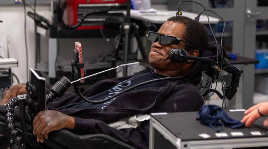
Brain-Computer Interfaces: A modern revolution in medicine and biomedical engineering
By: Dr Fizza Ali Tunio
Abstract
Brain–Computer Interfaces (BCIs) are innovative systems that translate neural activity into commands for external devices. By creating a direct connection between the brain and technology, BCIs are transforming modern medicine, particularly in rehabilitation, neuroprosthetics, communication systems, and neurological diagnostics. This article explores the neural foundations, brain regions involved, clinical applications, challenges, and future potential of BCIs in the medical field.
Introduction
Brain–Computer Interfaces represent a major advancement in biomedical engineering. These systems bypass conventional communication pathways by enabling direct interaction between the brain and external machines. Their development is driven by the need to assist individuals with neurological disabilities, enhance rehabilitation strategies, and expand the capabilities of medical diagnostics. BCIs integrate neuroscience, engineering, signal processing, and artificial intelligence into a unified, powerful technology.
Neural Foundations of BCIs
BCIs function by detecting electrical signals generated by neurons. Signals can be recorded using non-invasive methods, such as electroencephalography (EEG), or invasive approaches, such as implanted microelectrodes. Recorded neural data is processed through filtering, feature extraction, and machine-learning algorithms. The processed signals are then translated into specific commands, such as moving a robotic arm, controlling a wheelchair, or selecting letters on a communication interface.
Brain regions involved
3.1 Motor Cortex
The primary motor cortex (M1) encodes voluntary movements and is the core area for movement-based BCIs. Signals from this region allow users to control prosthetic limbs, robotic systems, and mobility aids.
3.2 Somatosensory Cortex
Adjacent to the motor cortex, the somatosensory cortex processes tactile and proprioceptive information. Including this region in BCIs enables sensory feedback, allowing users to “feel” pressure or touch through artificial limbs.
3.3 Visual Cortex
Located in the occipital lobe, the visual cortex is critical for visually driven BCIs, such as P300 or steady-state visually evoked potential (SSVEP) systems. These BCIs allow patients to select commands by focusing on specific visual stimuli.
3.4 Prefrontal Cortex
The prefrontal cortex is responsible for attention, decision-making, and executive functions. BCIs that utilize this area support cognitive monitoring and attention-based control.
3.5 Language Centers
Broca’s and Wernicke’s areas are fundamental for speech-related BCIs. Decoding neural patterns from these regions enables patients with speech impairments to communicate using synthesized language or text interfaces.
Medical Applications of BCIs
4.1 Neuroprosthetics and Motor Restoration
BCIs allow individuals with paralysis or motor impairments to control prosthetic limbs, exoskeletons, and assistive devices, significantly improving independence and quality of life.
4.2 Communication Systems
Patients with locked-in syndrome can communicate using BCIs that translate neural signals into text or speech, restoring interaction with caregivers and medical staff.
4.3 Rehabilitation Medicine
Motor-imagery BCIs help stroke patients and individuals with neurological injuries relearn movement by promoting neuroplasticity and reactivating damaged neural pathways.
4.4 Neurological Monitoring and Diagnostics
BCIs assist clinicians in monitoring epilepsy, assessing brain function during surgery, analyzing abnormal neural patterns, and evaluating consciousness and pain perception.
4.5 Support in Drug and Therapy Development
By observing brain responses to new drugs and therapies in real time, BCIs enable safer and more targeted pharmacological interventions for neurological disorders.
Challenges and ethical issues
Despite their promise, BCIs face several challenges:
- Signal stability – Neural signals can be noisy or inconsistent.
- Implant biocompatibility – Long-term invasive devices may cause tissue reactions.
- High cost – Advanced BCIs are expensive and require technical expertise.
- Training requirements – Users often need extended practice to operate devices efficiently.
Ethical considerations include neural data privacy, user autonomy, and informed consent, especially for vulnerable patients.
Future prospects
The future of BCIs is promising. Advancements in AI, wireless implants, and biocompatible materials are expected to:
- Enable fully integrated neuroprosthetic systems with sensory feedback
- Improve mental health therapy and cognitive rehabilitation
- Personalize medical treatments based on real-time neural activity
- Enhance human–machine interaction and expand neurotechnological applications
Conclusion
Brain–Computer Interfaces represent the convergence of neuroscience, engineering, and clinical medicine. By allowing the brain to interact directly with technology, BCIs offer unprecedented opportunities to restore lost functions, improve patient care, and transform modern medicine. As research continues, BCIs are poised to become an essential tool in rehabilitation, diagnostics, communication, and the development of advanced therapeutic strategies.
About The Author
Dr Fizza Ali Tunio is a fourth semester MBBS student at International University of Kyrgyzstan (IUK-ISM), Bishkek.

Advertisement
E-PAPER

Trending
Popular
Pakistan’s Dr. Misha Tanveer takes air pollution research to Europe ...
-
Calorie deficit for weight loss: How to ...
06:00 PM, 24 Aug, 2026 -
Vaccine maker warns of wider TBE, ...
05:00 PM, 21 Aug, 2026 -
FDA authorizes first robot to draw ...
06:11 PM, 20 Aug, 2026 -
One blood test for multiple cancers ...
04:52 PM, 11 Aug, 2026
Follow us on Facebook
Follow us on Twitter
Copyright ©2026 Medical News. All Rights Reserved.